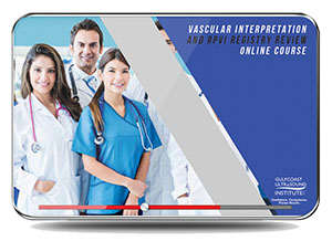
Vascular Interpretation & RPVI Registry Review
- A variety of learning components including: video lectures, scan demonstrations, interactive components/quizzes, and more to improve diagnostic accuracy.
- All online courses cover the same topics as our comprehensive live courses.
OVERVIEW
Vascular Interpretation & RPVI Registry Review Online Course is designed to provide a comprehensive review of the information needed to successfully pass the Physicians’ Vascular Interpretation (PVI) registry examination and to enhance vascular interpretation skills. The online activity is in a modular, interactive format and includes lectures, over 140 case presentations, and four mock examinations to simulate the certification exam experience. Enrollment is valid for 12 months (365 days) and begins the moment the participant is enrolled.
Additionally, the GCUS Registry Review PASS Guarantee offers you access to the online course version of your registry review program for an additional 90 days if you do not pass your registry exam after completing one of our review programs. Just send us a copy of your exam score (registry exam must be taken within six months of our review) and we’ll grant you a 90 day access extension to the online course version of the review.
OBJECTIVES
- Increase the participants’ knowledge and competence to perform/ interpret vascular ultrasound examinations and/or successfully take the RPVI certification exam.
- Analyze Doppler/Color physics factors that affect optimal Doppler examinations and commonly seen Doppler/Color artifacts, which may affect diagnostic accuracy.
- Recognize normal/abnormal imaging, spectral Doppler and Color Doppler findings seen with carotid, peripheral arterial, and venous disease.
- Cite the role of indirect testing in the evaluation of lower extremity arterial disease.
- Apply diagnostic criteria for evaluation of carotid, peripheral venous and arterial disease.
- Interpret complex carotid and peripheral vascular case studies in an interactive interpretation session format.
- Recognize normal/abnormal imaging, spectral Doppler and Color Doppler findings seen with Abdominal visceral (renal, mesenteric, and hepatic) evaluations.
- Recognize ultrasound findings associated with aortic endograft leaks.
- Differentiate normal and abnormal spectral Doppler characteristics associated with dialysis access graft evaluations.
- Integrate the key diagnostic elements into a structured report.
- Increase confidence to incorporate protocols, techniques, and diagnostic criteria to improve diagnosis/treatment accuracy.
AUDIENCE
TOPICS
- Hemodynamic Principles and Doppler Fundamentals
- Instrumentation: Transducers, Imaging Modes, and Artifacts
- Normal Carotid Anatomy, Waveforms, and Scan Protocol
- Abnormal Carotid Waveform: Characteristics & Diagnostic Criteria
- Abnormalities of the Vertebrobasilar System & TCD
- Integration of Data: Study Interpretation and Reporting
- Carotid and TCD Cases
- Abdominal Aorta and Aortic Endografts
- Peripheral Arterial: Indirect Physiologic Exams and Arterial Duplex/Color Flow Imaging
- Arterial Grafts and Intraoperative Duplex/Color Flow Imaging
- Arterial Assessment: Upper Extremities
- Abdominal Visceral Duplex: Renal, Mesenteric & Hepatic
- Peripheral Venous: Protocols and Methods to Detect DVT
- Venous Insufficiency and Physiologic Testing
- Venous Imaging: Upper Extremities
- Peripheral Arterial and Venous Cases
- Quality Assurance, Safety and Bioeffects
- Mock Exams
- Carotid Cases
- Peripheral Arterial Case Studies
- Abdominal Case Studies
- Miscellaneous Case Studies
Date of Original Release: 7/26/2021
This edition valid for credit through: 7/26/2024


Reviews
There are no reviews yet.