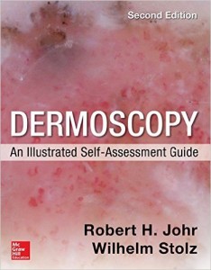
Dermoscopy An Illustrated Self-Assessment Guide 2e
by
Learn dermoscopy with this full-color, case-based self-assessment guide
With 436 clinical and dermoscopic images and 218 progressively more difficult cases commonly encountered in general dermatologic practice, Dermoscopy: An Illustrated Self-Assessment Guide offers a unique checklist methodology for learning how to use dermosocpy to diagnose benign and malignant pigmented and non-pigmented skin lesions.
Each high-quality, full-color clinical and dermoscopic image is presented with short history. Every case is followed by multiple-choice questions and three check boxes to test your knowledge of risk, diagnosis, and disposition. Turn the page, and the answers to the questions are provided in an easy-to-remember manner which includes the dermoscopic images being sown again. Circles, stars, boxes, and arrows appear in the image pointing out the important criteria of each case.
FEATURES:
- Cases involving the scalp, face, nose, ears, trunk and extremities, palms, soles, nails, and genitalia – many new to this edition
- The concepts of clinic-dermoscopic correlation, dermoscopic-pathologic correlation, and dermoscopic differential diagnosis are employed throughout
- Each case includes a discussion of all of its salient features in a quick-read outline style and ends with a series of dermoscopic and/or clinical pearls based on the authors’ years of experience
- Key dermoscopic principles are re-emphasized throughout the book to enhance your understanding and assimilation of the teaching points
- Two new chapters on trichoscopy and dermoscopy in general medicine
- Updated material on pediatric melanoma, desmoplastic melanoma, Merkel cell carcinoma, invasive squamous cell carcinoma, and nevi and melanoma associated with decorative tattoos
Product Details
- Paperback: 560 pages
- Publisher: McGraw-Hill Education / Medical; 2 edition (August 11, 2015)
- Language: English
- ISBN-10: 0071834346
- ISBN-13: 978-0071834346




Reviews
There are no reviews yet.