About This CME Teaching Activity
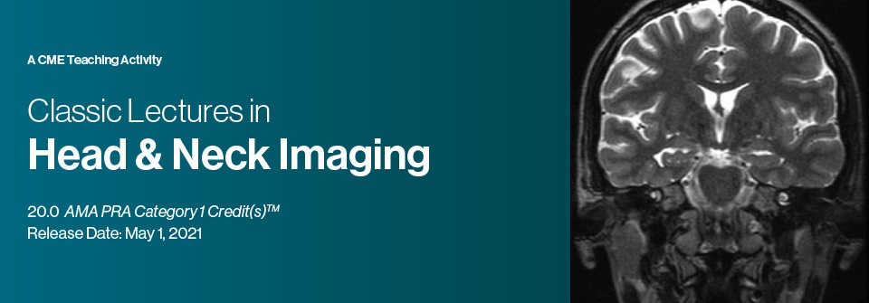
This CME activity brings together some of our most popular lectures in head & neck imaging. It combines a practical yet comprehensive review of head & neck imaging procedures concentrating on the latest trends, protocols and advances in clinical diagnosis, interpretation strategies and patient management. Faculty share techniques, tips and pitfalls through case based presentations.
Educational Objectives
At the completion of this CME activity, subscribers should be able to:
– Identify the anatomy of temporal bone, skull base, suprahyoid neck, and infrahyoid neck.
– Review anatomy and common pathology of the cranial nerves and brachial plexus.
– Identify the imaging features and clinical significance of perineural tumor spread.
– Review the anatomy, features and pathology of the lymphatic system.
– Recognize image features of lymphadenopathy.
– Describe the patterns of tumor spread associated with nasopharyngeal cancers.
– Determine the optimal imaging techniques for evaluating head and neck pathology.
– Describe obstacles associated with post-treatment imaging in head and neck cancer, and how to minimize them.
Target Audience
This CME activity should be of interest to radiologists, otolaryngologists, and referring physicians who order these types of studies.
Topics/Speakers:
Extramucosal Spaces of the Head and Neck
Laurie A. Loevner, M.D.
Lumps and Bumps: Central Topics in Head and Neck Radiology for the Non-subspecialist
Frank J. Lexa, M.D., MBA
Common Presenting Problems of the Head and Neck
C. Douglas Phillips, M.D., FACR
Orbital Anatomy and Pathology – Part 1
Gregg H. Zoarski, M.D.
Orbital Imaging
Frank J. Lexa, M.D., MBA
Orbital Anatomy and Pathology – Part 2
Gregg H. Zoarski, M.D.
Imaging of Orbital Tumors
C. Douglas Phillips, M.D., FACR
Orbital and Ocular Trauma: “More than Meets the Eye”
Alisa D. Gean, M.D.
Imaging Cranial Nerves I-VI
Blake A. Johnson, M.D., FACR
Cervical Lymphadenopathy
Richard H. Wiggins, III, M.D., CIIP, FSIIM
Imaging Cranial Nerves VII-XII
Blake A. Johnson, M.D., FACR
Brachial Plexus Imaging
Philip R. Chapman, M.D.
Imaging the Pituitary Gland and Sella Region
C. Douglas Phillips, M.D., FACR
Neuroimaging of Perineural Tumor Spread
Philip R. Chapman, M.D.
Imaging in Oropharyngeal and Oral Cavity Cancer
Lawrence E. Ginsberg, M.D.
Imaging of Laryngeal and Hypopharyngeal Cancer
Richard H. Wiggins, III, M.D., CIIP, FSIIM
Imaging Update in HPV-related Head & Neck Cancer
Lawrence E. Ginsberg, M.D.
Imaging and Biopsy Issues in Thyroid Malignancy
C. Douglas Phillips, M.D., FACR
Imaging Issues in Hyperparathyroidism Including 4D CT
Deborah R. Shatzkes, M.D.
Neuroimaging of the CPA, Internal Auditory Canal and Inner Ear
Philip R. Chapman, M.D.
Central Skull Base: Pathology and Anatomy
C. Douglas Phillips, M.D., FACR
Imaging of Salivary Neoplasia
Deborah R. Shatzkes, M.D.
Suprahyoid Neck: Spatial Approach
C. Douglas Phillips, M.D., FACR
Imaging of Cutaneous Malignancy
Lawrence E. Ginsberg, M.D.
Imaging the Patient with Hearing Loss
C. Douglas Phillips, M.D., FACR
MRI of the Temporal Bone
Wende N. Gibbs, M.D.
Imaging of Temporal Bone Neoplasia
C. Douglas Phillips, M.D., FACR
Sinonasal Infectious and Inflammatory Pathology
Richard H. Wiggins, III, M.D., CIIP, FSIIM
Imaging of Sinonasal and Anterior Cranial Base Lesions
Deborah R. Shatzkes, M.D.
Imaging of Nasopharyngeal Carcinoma
Richard H. Wiggins, III, M.D., CIIP, FSIIM
Radiation Therapy Treatment Planning for the Head and Neck: Emphasis on NPC
David I. Rosenthal, M.D.
Nasopharyngeal Cancer: Patterns of Spread
Laurie A. Loevner, M.D.
Top 10 Head and Neck Imaging Pearls
Richard H. Wiggins, III, M.D., CIIP, FSIIM
Case Based Review of Head and Neck Lesions
Laurie A. Loevner, M.D.
Note : The lecture Thyroid Nodules and Nodules that are Cancer is not available from Publisher
CME Release Date 1/05 /2021
CME Expiration Date 4/30/2024







Reviews
There are no reviews yet.