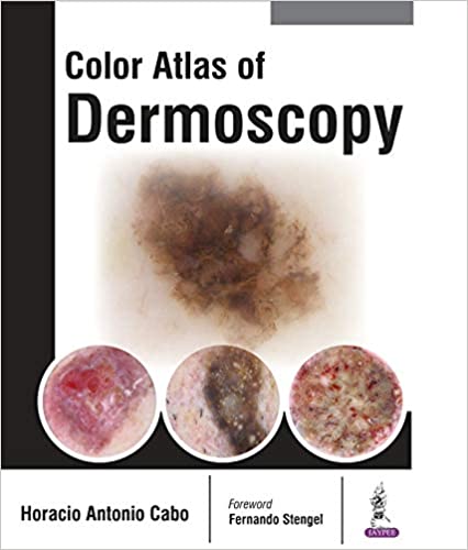
This book focuses on Color Atlas of Dermoscopy. Dermoscopy is a noninvasive method that allows the in vivo evaluation of colors and microstructures of the epidermis, the dermoepidermal junction, and the papillary dermis not visible to the naked eye. These structures are specifically correlated to histologic features. The identification of specific diagnostic patterns related to the distribution of colors and dermoscopy structures can better suggest a malignant or benign pigmented skin lesion. The use of this technique provides a valuable aid in diagnosing pigmented skin lesions. This book consists of 18 chapters, which include dermatoscope, structures, patterns, criteria and colors, vascular patterns, nonmelanocytic lesions, melanocytic lesions, melanoma simulators, combined lesions, special locations, diagnostic algorithms, total-body photography and sequential digital dermoscopy images, revised pattern analysis, entomodermoscopy, inflammatoscopy, trichoscopy, capillaroscopy, and reflectance confocal microscopy. The information in this state-of-the-art volume is presented in a simple manner, with the aid of clear diagrams that emphasize the things. Data are highlighted with the use of tables that single out the characteristic signs of each individual entity. This book is very useful for the readers to improve their dermoscopic skills for the benefit of their patients.
Product details
- Item Weight : 1.98 pounds
- Hardcover : 400 pages
- ISBN-10 : 9386056305
- ISBN-13 : 978-9386056306
- Product Dimensions : 8.5 x 1 x 10.5 inches
- Publisher : Jaypee Brothers Medical Pub; 1st Edition (July 17, 2017)
- Language: : English


Reviews
There are no reviews yet.